 Skip to content

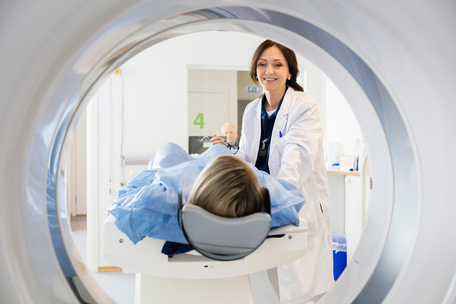
CT scans and MRI scans can both create detailed images of what is happening inside the body, but they use very different technologies and are often chosen for different medical reasons.
A CT scan uses X-rays and computer processing to create cross-sectional images of organs, bones, blood vessels, and other structures. MRI uses a powerful magnetic field and radio waves instead of ionizing radiation. Both can provide valuable diagnostic information, but one is not automatically better than the other. The right choice depends on the part of the body being examined, the condition your physician is evaluating, how quickly results are needed, and your individual medical history.
At Impact Health NY, imaging is selected based on clinical indication rather than simply ordering the most advanced test available. Physician interpretation matters because the most useful scan is the one that answers the right medical question.
What Is a CT Scan?
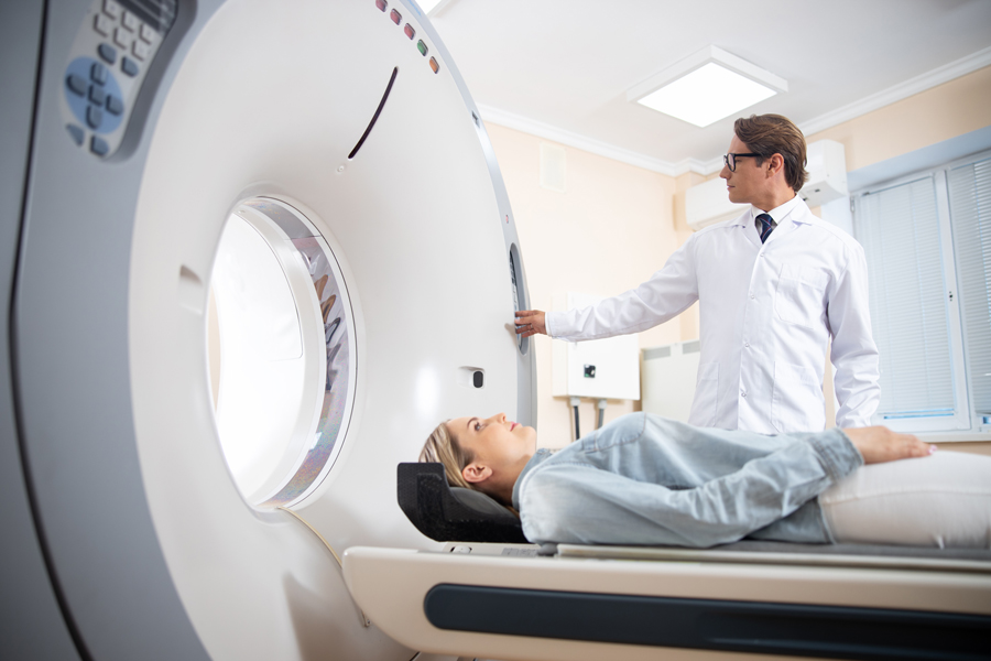
CT stands for computed tomography. A CT scanner uses X-rays taken from multiple angles and computer processing to create detailed cross-sectional images of the body.
CT can show bones, organs, soft tissues, and blood vessels and can reconstruct images in multiple planes or three dimensions. CT scans are also relatively fast, which makes them especially useful when physicians need detailed information quickly.
Depending on the reason for the scan, contrast material may be used to make certain organs, blood vessels, or abnormalities easier to see.
CT imaging is commonly used to evaluate:
- The lungs and chest
- The heart and coronary arteries
- Bones and fractures
- Internal injuries or bleeding
- Abdominal and pelvic concerns
- Blood vessels
- Certain tumors and other abnormal findings
The exact type of CT scan depends on the medical concern being investigated.
What Is an MRI?
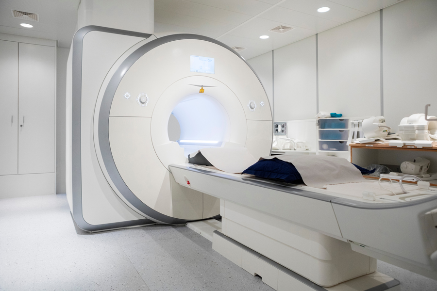
MRI stands for magnetic resonance imaging. Instead of X-rays, MRI uses a strong magnetic field, radio waves, and computer processing to produce detailed images of internal structures. MRI does not use ionizing radiation.
MRI is particularly useful when physicians need detailed views of many types of soft tissue.
It can be used to evaluate areas such as:
- The brain and spinal cord
- Muscles, tendons, and ligaments
- Joints
- The heart
- Blood vessels
- The liver and other abdominal organs
- Certain tumors or abnormal tissue
Some MRI studies use a gadolinium-based contrast agent to provide additional detail.
CT Scan vs. MRI: What Is the Main Difference?
The biggest difference is how the images are created.
A CT scan uses ionizing X-rays. MRI uses magnetic fields and radio waves.
That difference affects how quickly the scan can be performed, what types of tissue it shows particularly well, what safety considerations apply, and whether radiation exposure is involved.
CT tends to be faster and is excellent for evaluating many problems involving the lungs, bones, blood vessels, and acute injuries. MRI often provides greater soft-tissue detail and is especially useful for the brain, spinal cord, joints, muscles, and selected cardiac or abdominal conditions.
Neither test replaces the other in every situation.
Is a CT Scan Faster Than an MRI?
Usually, yes.
CT scans can often collect the necessary images within minutes. This speed can be especially valuable in emergency situations, such as suspected internal bleeding, serious injury, or other conditions where rapid diagnosis matters.
An MRI scan usually requires more time as it may take several minutes for the patient to stay still inside the machine.
It can also help make the procedure less difficult for the patient due to its shorter duration.
Which Scan Provides More Detail?
That depends on what your physician is looking for.
MRI often provides greater contrast between different types of soft tissue. This can make it particularly useful for evaluating the brain, spinal cord, ligaments, tendons, muscles, and certain internal organs.
CT gives great anatomical details and is capable of showing bones, lungs, blood vessels, soft tissue, and internal organs all at once. It is also great for several cardiovascular and pulmonary studies.
Comparing MRI with CT based on which one gives better detail can thus be confusing.
CT vs. MRI for Heart Imaging
Both CT and MRI can play important roles in cardiovascular care, but they answer different questions.
Cardiac CT can provide detailed images of the coronary arteries and is used for tests such as coronary artery calcium scoring and coronary CT angiography.
A coronary calcium score looks for calcified plaque in the coronary arteries. Coronary CT angiography, or CCTA, uses intravenous contrast to provide a more detailed view of the arteries and can help identify plaque and narrowing.
Cardiac MRI is commonly used when physicians need detailed information about the heart muscle, heart structure, tissue characteristics, blood flow, or certain forms of heart disease.
At Impact Health NY, cardiovascular imaging is chosen based on symptoms, medical history, previous testing, risk factors, and the specific information needed to guide care.
Does a CT Scan Use Radiation?
Yes. CT uses ionizing radiation.
The radiation dose varies depending on the type of scan, the body area being examined, the equipment, and the imaging protocol. The FDA notes that when a CT scan is medically appropriate, its diagnostic benefit generally outweighs the relatively small radiation risk. Unnecessary exposure should still be avoided.
This is one reason Impact Health NY takes a selective approach to imaging. More scans do not automatically lead to better preventive care.
Does an MRI Use Radiation?
No. MRI does not use ionizing radiation.
Instead, it relies on strong magnetic fields and radiofrequency energy. That makes MRI useful when avoiding radiation is particularly important, but the magnetic environment introduces its own safety considerations.
Patients with implants, electronic devices, metal fragments, or other medical hardware need to discuss them with the imaging team before entering an MRI scanner.
CT Contrast vs. MRI Contrast
Both CT and MRI can be performed with or without contrast, depending on the examination.
CT commonly uses iodine-based contrast material. Contrast can make blood vessels, organs, and certain abnormalities easier to distinguish. Patients with previous contrast reactions or significant kidney disease may require additional review before receiving it.
MRI typically uses gadolinium-based contrast when contrast is needed. Gadolinium is different from the iodine-based contrast used for CT. Patients with kidney disease, previous gadolinium reactions, or certain other health conditions should discuss them with the imaging team beforehand.
Not every CT or MRI requires contrast.
What Are the Advantages of a CT Scan?
One of CT’s biggest advantages is speed.
A CT scan can rapidly provide detailed images of several types of tissue at once, including bone, organs, and blood vessels. It is also less sensitive to patient movement than MRI.
CT can be particularly useful when:
- Results are needed quickly
- The lungs need to be evaluated
- Bone injury is suspected
- Internal bleeding or trauma is a concern
- Coronary arteries or other blood vessels need detailed imaging
- A physician needs clear anatomical information
CT is also generally easier to perform in patients who have difficulty remaining still for long periods.
What Are the Advantages of an MRI?
MRI provides detailed soft-tissue imaging without ionizing radiation.
That can make it particularly useful when physicians are evaluating:
- The brain
- Spinal cord
- Ligaments and tendons
- Muscles
- Joints
- Certain heart conditions
- Selected abdominal or pelvic abnormalities
MRI can also provide information about tissue characteristics that may not be as clear on CT.
The tradeoff is that MRI usually takes longer and requires careful screening for metallic or electronic implants.
What Are the Limitations of CT?
The main consideration is radiation exposure.
CT may also require iodine-based contrast, which can cause reactions in some patients and may require kidney function to be reviewed.
Another consideration is incidental findings. Because CT scans provide detailed images, they can sometimes identify unrelated abnormalities that require additional evaluation. The FDA notes that incidental findings can lead to further testing, even when the finding ultimately proves harmless.
CT also does not provide the same soft-tissue characterization as MRI for certain conditions.
What Are the Limitations of MRI?
MRI can take longer and generally requires the patient to remain very still.
Some people also find the enclosed scanner uncomfortable or experience claustrophobia.
The strong magnetic environment requires careful review of pacemakers, cochlear implants, pumps, metal fragments, and other implanted or external medical devices. Some devices are MRI-safe or MRI-conditional, while others are not suitable for the MRI environment.
MRI can also be more sensitive to patient movement, which can reduce image quality.
Can You Have a CT Scan With a Pacemaker or Implant?
In many cases, yes.
CT generally creates fewer concerns for patients with implanted medical devices than MRI. The FDA notes that CT can often be performed safely in patients with electronic medical devices, although appropriate precautions may still be necessary.
MRI requires more careful device screening. Some modern pacemakers and implants are MRI-conditional, meaning they can be scanned under specific conditions. Devices with unknown MRI safety status require additional evaluation before MRI is performed.
Always tell the imaging team about any implanted or wearable medical device before either test.
How Do Doctors Decide Between CT and MRI?
The decision starts with the clinical question.
A physician may consider:
- Your symptoms
- The body area being examined
- Your medical history
- Previous imaging results
- How quickly information is needed
- Whether blood vessels need to be evaluated
- Whether radiation exposure is a concern
- Kidney function
- Previous contrast reactions
- Pregnancy
- Implanted devices or metal
- Whether the result is likely to change your treatment or prevention plan
Sometimes CT is clearly the better choice. In other situations, MRI offers information that CT cannot provide as effectively. Occasionally, both tests may be used because they answer different questions.
Is One Safer Than the Other?
Neither CT nor MRI is universally safer.
MRI avoids ionizing radiation, which is an important advantage. However, its strong magnetic field creates safety considerations involving certain implants, metal objects, and electronic devices.
CT involves radiation, but for a medically indicated scan, the potential diagnostic benefit generally outweighs the small associated risk.
The safest approach is not simply choosing the test with fewer theoretical risks. It is choosing the test most appropriate for the patient and the medical question.
Choose the Right Imaging With Physician Guidance
The right imaging test should answer a specific medical question and help guide your next steps. At Impact Health NY, Dr. Marcus considers your medical history, symptoms, laboratory findings, genetics when relevant, and individual risk factors before recommending CT or other advanced imaging.
If you are unsure which imaging test may be appropriate, schedule a consultation today. We can review your health concerns, discuss your options, and determine whether advanced imaging has a meaningful role in your preventive care.
CT and MRI FAQs
Is an MRI better than a CT scan?
Not necessarily. MRI often provides greater soft-tissue detail, while CT is faster and can be particularly useful for evaluating bones, lungs, blood vessels, and certain cardiovascular conditions. The better test depends on the reason for imaging.
Which scan takes longer, CT or MRI?
MRI generally takes longer. CT image collection can often be completed within minutes, while an MRI may require a longer period of scanning.
Which scan uses radiation?
CT uses ionizing radiation. MRI does not.
Can both CT and MRI use contrast?
Yes. Some CT studies use iodine-based contrast, while some MRI studies use gadolinium-based contrast. Many examinations can also be performed without contrast.
Which test is better for the heart?
That depends on what needs to be evaluated. CT can provide detailed views of coronary calcium, coronary plaque, and artery narrowing. MRI can provide detailed information about heart muscle, structure, function, and tissue characteristics.
Can you get an MRI if you have metal in your body?
Sometimes. Many modern implants are MRI-safe or MRI-conditional, but others may not be suitable. The exact device needs to be identified and reviewed before the scan.
Why would a doctor order a CT instead of an MRI?
CT may be preferred when rapid imaging is needed or when the lungs, bones, coronary arteries, blood vessels, internal bleeding, or certain other conditions need to be evaluated.
How do I know which scan I need?
The right imaging study depends on your symptoms, medical history, risk factors, and the question your physician needs the scan to answer. A medical evaluation should guide the choice rather than selecting an imaging test on your own.